X-ray microscopy
Soft x-rays are a form of high-energy electromagnetic radiation with wavelengths ranging from 0. 1 to 10 nm. Because of their short wavelength, x-rays offer penetration depths unparalleled to any other imaging technique.
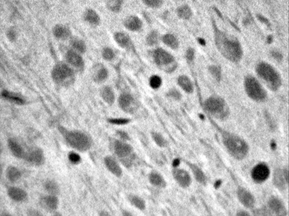
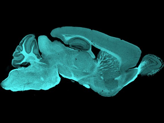
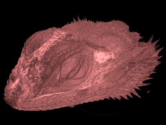
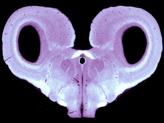
X-ray computed tomography (CT) relies on two processes. The photoelectric absorption of the x-rays by matter, which creates cross-sections of physical objects, and tomographic reconstruction algorithms, which produce 3D volume renderings from two-dimensional images. X-ray micro-CT microscopes can image unstained soft tissues and perform virtual histology on a very wide range of samples, from small brain sections to whole adult animals.
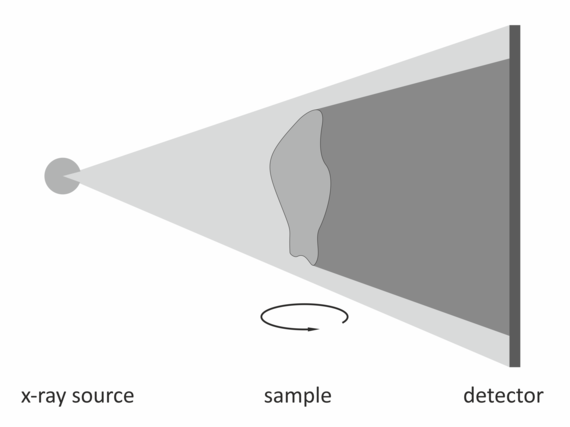
Because x-rays have very small wavelengths, bending and reflecting them is virtually impossible. Hence, ever since Wilhelm Röntgen's first x-ray in 1895, the way images are taken has not changed. Any typical x-ray micro-CT consists of an x-ray source, the sample and a detector (see schematic below). The positions of the source and detector with respect to the sample determine the magnification of the image. The rule of thumb is the closer the sample to the source and the further the sample, the higher the magnification.
We provide support in sample preparation as well as access, training and project support on our Zeiss XRadia 520 Versa.
Bobtail squid
Whole bobtail squid (Euprymna berryi), stained with lugol solution.
Staining and sample preparation by David Hain, imaging by Claudio Polisseni
