Lightsheet microscope with 2-photon excitation
Custom lightsheet with even sheet-thickness across a large field of view
Though confocal microscopes are the workhorses of most fluorescence microscopy-based research facilities, they have a few disadvantages. Compared to widefield fluorescence microscopy
- the acquisition speed is very slow, since only one pixel at a time is imaged
- phototoxicity and bleaching are very strong if 3D volumes are acquired
In standard light-sheet microscopy, a very thin focused slice of light is used for the excitation of the fluorophores in the sample. The fluorescence light is imaged with a second objective, often orthogonally oriented.
This way only the imaged volume is illuminated, which is reducing bleaching and phototoxicity a lot. The acquisition is done with a camera facilitating highly parallelized readout and a huge increase in acquisition speed. Modern sCMOS cameras offer a three times higher quantum efficiency compared to a standard confocal detector, increasing both acquisition speed and signal to noise ratio.
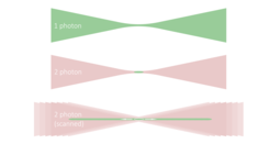
The achievable resolution is usually slightly reduced when compared to a very good confocal microscope though. It is particularly compromised in the regions outside the lightsheet focus, where the lightsheet becomes thicker due to the divergence of the light.
For having the best of both worlds, we developed a custom light-sheet setup ("Santoku") that uses 2-photon excitation for reducing scattering in the sample for deeper penetration. By axially scanning the light-sheet across a large FOV of 1.2 mm x 1.2 mm we achieve a homogenous lightsheet with excellent axial resolution across the whole scanning range. The Santoku microscope supports refractive indices between 1.33 (water) and 1.55 (Cubic and iDISCO) and has a resolution of around 0.6 µm x 0.6 µm x 3 µm, while supporting samples up to mouse brain size. A whole mouse brain can be imaged in roughly 2 hours (depending on the resolution).

Middle: two-photon excitation in a focussed laser beam. Excitation is limited to the region surrounding the focus.
Bottom: If the focus is scanned along the optical axis a long uniform region is excited which does not suffer from the divergence found in the case of one-photon excitation


