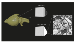
Heavy-metal staining for electron microscopy
The imaging technique to investigate the connectome - the connections between neurons - is electron microscopy (EM). This technique however requires drastic staining techniques, based on heavy metals like osmium. Hua and coworkers from the Department of Connectomics at the Max Planck Institute for Brain Research investigated the mechanisms for osmium staining. Based on these new insights the researchers designed a new staining protocol, which makes staining of neurons for EM almost a trivial enterprise. They published their findings in the latest edition of Nature Communications.

New methods to efficiently stain neurons for EM
In order to visualize brain circuits with electron microscopy, drastic staining with heavy metals, like osmium and uranium. Although, these methods are obviously not fully optimized, the basic procedures were not changed for decades.
In the Nature Communications paper, Hua et al describe a method to optimize the balance between an optimal attachment to the membranes and the diffusion. By studying the different valencies of the osmium compounds at the different reaction steps, the researchers could design a protocol, where they could efficiently balance the diffusion and attachment steps.
Helmstaedter: "This new, almost trivial, protocol was successfully tested on mouse tissue from the neocortex and gives excellent staining results. Possibly, it can also be further investigated for other uranium-based staining methods and to even larger tissues".


