New insights in Autism Spectrum Disorders via abstract face recognition
Magneto-encephalography (MEG) was used to show differences in visual processing between adults with Autism Spectrum Disorders and the control group.
Using Mooney face stimuli, Peter Uhlhaas and colleagues from the Max Planck Institute for Brain Research and the Ernst Strüngmann Institute as well as researchers from the Goethe University in Frankfurt am Main showed lower activity in the gamma-band for adults with Autism Spectrum Disorders. Additionally, they found that both groups process the task in different brain regions. Their results were published in the latest issue of the Journal of Neuroscience.

Autism Spectrum Disorders (ASD) are associated with non-typical social interaction, communication and perception, as well as higher level cognition but the underlying neurobiological abnormalities are unclear. In this study, magneto-encephalography (MEG), a non-invasive brain imaging technique was used, which allows the measurement of neuronal activity on a millisecond time-scale. The researchers focused on a particular frequency range of neuronal oscillations in the brain; the gamma band (between 30 and 200 Hz), believed to be important for coordinated brain activity.
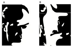
Peter Uhlhaas: "We obtained MEG-data with a group of adult participants with ASD and a control group while they had to perform a Mooney face stimuli recognition task. During this task, all participants had to detect a face in a complex display of two types of black and white drawings (A and B, see Figure). Participants with ASD were particularly impaired when they had to group the stimulus elements into a meaningful facial representations (A) which could underlie impairments in social communication."
An analysis of the MEG data showed reduced power of gamma-band oscillations during face perception in the ASD group which indexes impaired temporal precision of cortical circuits. Moreover, participants with ASD also activated different brain areas than the control group. While in controls, gamma-band activity was found in regions which are important for assigning meaning and emotions to facial stimuli, participants with ASD showed the strongest activation in brain regions which are responsible for interpreting simple objects.
Uhlhaas: "These results show that participants with ASD have a pronounced impairment in coordinating brain activity appropriately which leads to different task-strategies. These results may also be important in understanding the biological mechanisms of ASD because we know that specific neurotransmitter-systems are involved in generating high-frequency oscillations during normal brain functioning which could be impaired in the disorder"
Contact: Dr. Peter J. Uhlhaas, Department of Neurophysiology, Max Planck Institute for Brain Research, Deutschordenstr. 46, 60528 Frankfurt am Main, Germany, Email: peter.uhlhaas@brain.mpg.de